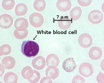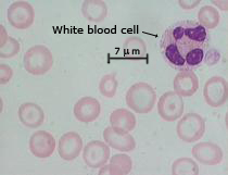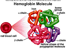
Figure 4-1. Hemoglobin consists of 2 β-globin proteins and 2 α-globin proteins. (Click image on right for larger version.) Illustration used with permission from McGraw-Hill. Sylvia S. Mader, Inquiry into Life, 8th ed. © 1997 The McGraw-Hill Companies, Inc. All rights reserved.
The hemoglobin forms a lattice structure on the cytoplasmic side of the RBC plasma membrane; thus, affecting the shape of the RBC, as shown in Panels A, B, and C of Figure 4-2 below.
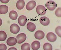
Figure 4-2. Three different RBC samples. The iron-containing heme group is pigmented. Cells with less heme have less pigmentation and are said to be hypochromic. Note: mature RBC lack a nucleus. Also note: The following image location is the reference for the 7 micron size of a normal red blood cell: http://static.howstuffworks.com/gif/artificial-blood-7.jpg
![]() |
![]() |
![]() |
Panel A: Normal sample | Panel B: Mrs. Williams’ sample | Panel C: Tazswana’s sample |
Credit: Copyright © The American Society for Clinical Laboratory Science. Used with permission. | Credit: Donald Innes, M.D. Used with permission. | Credit: Donald Innes, M.D. Used with permission. |
1. Compare the size, shape and color intensity of the red blood cells. Record your observations in the chart below:
![]() | ![]() | |||
---|---|---|---|---|
Panel A: Wild-type | Panel B: Mrs. Williams | Panel C: Tazswana | ||
Size | ||||
Shape | ||||
Color Intensity |
2. Thinking about the function of β-globin and the shape and color of Tazswana’s red blood cells, explain why Tazswana has severe symptoms compared to her mother or a normal person.
Originally published at http://www.sciencecases.org/tazswana/tazswana4.asp
Copyright © 1999–2025 by the National Center for Case Study Teaching in Science. Please see our usage guidelines, which outline our policy concerning permissible reproduction of this work.